A blog celebrating the the many cycles in life, nature, science, and nature.
Read MoreAurora Borealis, Yukon, Canada. © Joseph Bradley / Science Source

Aurora Borealis, Yukon, Canada. © Joseph Bradley / Science Source
A blog celebrating the the many cycles in life, nature, science, and nature.
Read More
Measles virus particle, illustration. © Kateryna Kon / Science Source
The first written description of measles appeared in the 9th century, recorded by Persian physician Muhammad ibn Zakariya al-Razi.
This highly contagious viral infection originated from a mutation of a bovine disease called rinderpest. Through global vaccination efforts, rinderpest was eradicated in 2011.
While cattle are now safe, humans remain vulnerable to the measles virus.
With global travel, measles spread rapidly across continents, leaving illness and death in its wake.
Those infected typically develop a high fever, cold-like symptoms, and a rash. Most recover within a few weeks, but some develop pneumonia or encephalitis. Pregnant women face the risk of severe complications, including harm to their unborn child.
The worst U.S. outbreak occurred before the vaccine was developed, causing nearly 500 deaths and over half a million infections.
The measles vaccine, developed in 1963 and widely available by 1971, is 97% effective. Yet some still put children—and others—at risk by avoiding vaccination. Even a mild case can transmit the virus to infants, older adults, or those with weakened immune systems, leading to serious illness or death.
In rare cases, measles can lead to a fatal complication known as subacute sclerosing panencephalitis (SSPE).
Everyone has the choice to vaccinate, but it’s vital that decisions are based on accurate medical information.
Science Source provides authentic medical imagery, illustrations, and infographics to support your healthcare communication projects.

Measles Illustration
Symptoms, complications and infections rate. © Monica Schroeder / Science Source

Baby with Measles Rash
A baby infected with the Morbillivirus, the cause of measles, or rubeola. © Betty Partin / Science Source

TEM of Measles (rubeola) Virus
Colored transmission electron micrograph (TEM) of a measles virus (rubeola), with its envelope broken exposing the nucleocapsid filaments. © Dr. Linda Stannard / Science Source

Rinderpest Plague, 1868
Cattle dying in the slaughterhouse yards, during the cattle plague. Measles developed from a mutation of the rinderpest virus. © NLM / Science Source

Spinosaurus aegyptiacus in its paleo-environment: North Africa (Egypt) around 95 million years ago. This Spinosaurus is a male in its breeding colouration; we can see this from the brightly colored throat and the yellow blotches on the sail. This male is patrolling its territory in search of a potential partner. © Simone Zoccante / Science Source
Science Source is excited to welcome three new paleoartists—Simone Zoccante, Gustavo Higón, and Mohamad Haghani to our collection. Each brings prehistoric creatures to life while recreating Earth’s ancient landscapes with the latest scientific insights.
Simone Zoccante, an Italian artist with nearly a decade of experience, has become a leading figure in contemporary paleoart. His crisp, highly detailed digital style merges scientific accuracy with a striking modern look. Featured here is his Spinosaurus, a dinosaur built for aquatic hunting, with a crocodile-like tail for propulsion, dense bones that kept it submerged, and a sail that may have aided swimming or served as display. Altogether, these traits made it one of the best swimmers of land-dwelling dinosaurs.
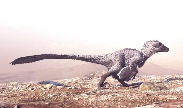
Computer illustration depicting an adult Dakotaraptor steini. © Gustavo Higón/Science Source
Gustavo Higón, based in Spain, is known for his cinematic paleoart. His illustrations often place dinosaurs in stark, desert environments, capturing both their power and fragility. His illustration of an adult Dakotaraptor steini in full stride shows one of the largest raptors —over 16 feet long, feathered, and built for speed. The scene emphasizes its anatomy and locomotion, illustrating how this predator hunted and explored its ecosystem.

Two Elasmosauruses swimming through the water, chasing and preying upon schools of fish in a dynamic underwater scene. © Mohamad Haghani / Science Source
Mohamad Haghani, from Iran, has been working in paleoart for nearly ten years. Combining detailed anatomy with carefully reconstructed environments, Haghani conveys the most current scientific research with striking, realistic imagery.
His two Elasmosauruses swimming through the water, captures the long necks, paddled limbs, and slender bodies of the species. Using current research, he highlights how they maintained a straightened posture and an out-stretched neck while hunting.
Together, these artists expand our growing collection of paleoart. Explore their work and many other illustrators and photographers covering prehistoric life, fossils, and more at ScienceSource.com.

Microscopic photo of Sickle cells, crescent-shaped erythrocytes (red blood cells) that result from a change in the amino acid sequence of the cells' hemoglobin. © Eye of Science / Science Source
Sickle cell disease (SCD) is one of the world's most common inherited blood disorders, affecting millions of people globally. In the United States, approximately 100,000 individuals live with the disease, with sickle cell disease occurring in about 1 in 365 Black or African American births. The condition is also prevalent in regions where malaria has historically been common, including sub-Saharan Africa, the Middle East, India, and parts of the Mediterranean.

Sickle Cell Patient in Clinic
A nurse connecting tubes to a SCD patient's arm for a red blood cell exchange. © Life in View/Science Source
Caused by a genetic mutation in hemoglobin, sickle cell disease transforms normally flexible red blood cells into rigid, crescent-shaped cells that can block blood flow and reduce oxygen delivery throughout the body. These blockages can lead to severe pain crises, anemia, infections, organ damage, stroke, and other life-threatening complications.
Recent advances in treatment have transformed patient care. New medications, improved supportive therapies, stem cell transplantation, and emerging gene-editing approaches offer hope for improved outcomes and, in some cases, a potential cure. Yet many patients continue to face significant health challenges, disparities in care, and limited access to advanced treatments.
As researchers continue to explore innovative therapies, clear and accurate communication remains essential for patients, healthcare professionals, educators, and the public.

Genetics of Sickle Cell Disease
Chromosome 11, a normal blood cell, and a sickle cell. © Monica Schroeder/Science Source
Sickle cell disease involves complex biological processes that can be difficult to explain through text alone. Authentic medical imagery helps audiences understand abnormal red blood cell morphology, vaso-occlusion, genetic inheritance, disease complications, diagnostic testing, and emerging treatments.
High quality visuals can improve engagement and comprehension in textbooks, patient education materials, healthcare marketing campaigns, pharmaceutical communications, news stories, and digital health platforms.
Science Source provides a comprehensive collection of authentic, expertly captioned medical stock photography, illustrations, micrographs, and medical imaging related to sickle cell disease. Our content is created by medical photographers, scientific illustrators, and healthcare professionals, not generated by AI, ensuring the accuracy and credibility required for professional healthcare and educational use.
Gallery of Sickle Cell Disease Images for Licensing
Whether you are developing educational resources, reporting on advances in gene therapy, creating patient-facing materials, or illustrating scientific publications, Science Source offers trusted visual content that helps audiences better understand this complex genetic disorder.

3D Illustration of Blood Clot
Sickle cells cannot move through small blood vessels causing blockages. © Tim Vernon/Science Source

Link Between Sickle Cell and Strokes
Sickle cell patients are more prone to stroke. © Sue Seif/Science Source